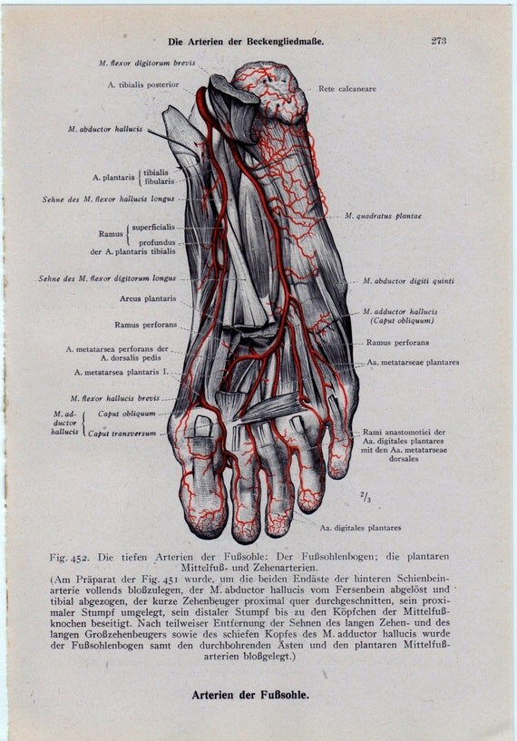
Anatomical Foot Diagram
View Anatomical Foot Diagram Images. We hope this picture foot external anatomical landmark and internal view can help you study and research. Two longitudinal (medial and lateral) arches and one anterior transverse arch.

Our foot and ankle chart is one of our best selling charts, perfect for learning and explaining the major bony features of the foot and ankle.
They are formed by the tarsal and metatarsal bones, and supported by ligaments and tendons in. However, they may also eventually represent a source of pathology, such as painful syndromes. Foot and leg anatomy these look like netters plates. We hope this picture foot external anatomical landmark and internal view can help you study and research.
0 Response to "Anatomical Foot Diagram"
Post a Comment